The superior oblique muscle is one of the six extraocular muscles that control movement of the eye. Although it is a relatively small muscle, its unusual course through a pulley-like structure called the trochlea gives it an important role in eye rotation and downward movement.
In simple terms, the superior oblique helps the eye look downward and outward and rotate inward. It is also unique because it is the only extraocular muscle supplied by the trochlear nerve (cranial nerve IV).
Understanding this muscle is particularly important when studying eye anatomy, cranial nerves, diplopia, strabismus, and conditions such as superior oblique palsy, Brown syndrome, and superior oblique myokymia.
What Is the Superior Oblique Muscle?
The superior oblique is one of the six muscles that move the eyeball:
- Superior rectus
- Inferior rectus
- Medial rectus
- Lateral rectus
- Superior oblique
- Inferior oblique
The superior oblique is located in the upper medial portion of the orbit. Unlike the four rectus muscles, it does not simply run directly from the back of the orbit to the eyeball.
Instead, it travels forward toward the medial upper part of the orbit, passes through the trochlea, and then changes direction before attaching to the back and outer-upper portion of the eyeball.
This pulley-like arrangement is the key to understanding why the muscle can produce its characteristic eye movements.
Quick facts
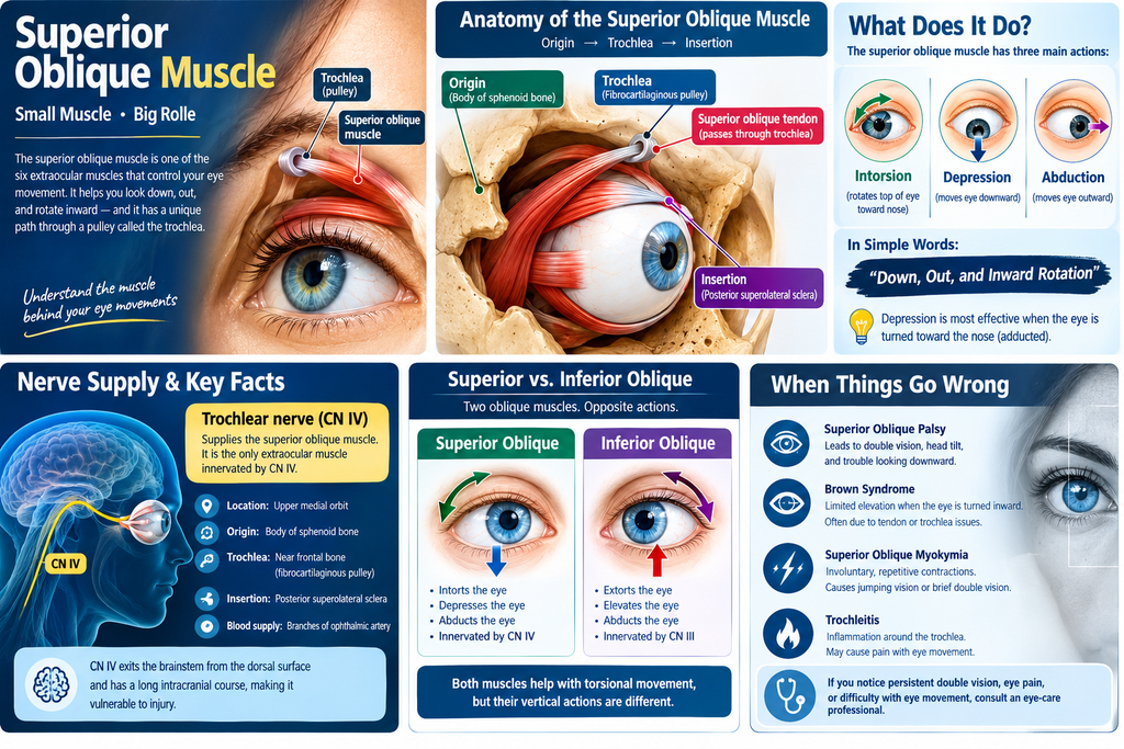
| Feature | Superior oblique muscle |
|---|---|
| Location | Upper medial orbit |
| Origin | Body of the sphenoid bone |
| Trochlea | Fibrocartilaginous pulley near the frontal bone |
| Insertion | Posterior superolateral sclera |
| Main action | Intorsion |
| Other actions | Depression and abduction |
| Nerve supply | Trochlear nerve (CN IV) |
| Blood supply | Branches of the ophthalmic artery |
The exact anatomical dimensions and relationships can vary between individuals.
Superior Oblique Muscle Origin and Insertion
The origin of the superior oblique is on the body of the sphenoid bone, near and medial to the optic canal.
From there, the muscle runs forward along the medial orbital wall. Near the front of the orbit, it becomes a tendon and passes through the trochlea.
The trochlea acts much like a pulley. After passing through it, the tendon changes direction and travels posterolaterally beneath the superior rectus muscle. It eventually attaches to the sclera in the posterior superolateral region of the eyeball.
This unusual pathway is one of the most important anatomical features of the superior oblique.
Why is the trochlea important?
The trochlea changes the direction in which the muscle pulls on the eyeball.
Without this pulley-like redirection, the superior oblique would not produce its characteristic combination of torsional, vertical, and horizontal movements.
The relationship between the muscle, tendon, and trochlea is also clinically important because abnormalities affecting this complex can interfere with normal eye movement.
What Does the Superior Oblique Muscle Do?
The superior oblique has three principal actions:
- Intorsion – rotates the top of the eye toward the nose.
- Depression – moves the eye downward.
- Abduction – moves the eye outward.
Its primary action in the primary position is intorsion, while depression and abduction are secondary and tertiary actions.
A useful way to remember its movement is:
Superior oblique = down, out, and inward rotation.
However, the muscle does not perform these movements equally in every position of gaze. Its mechanical effect changes depending on where the eye is looking.
When the eye is adducted
The superior oblique is particularly effective as a depressor when the eye is turned toward the nose.
This is clinically important because weakness of the superior oblique can cause difficulty controlling vertical alignment when looking downward.
Superior Oblique Muscle Innervation
The superior oblique muscle is supplied by the trochlear nerve (cranial nerve IV).
This is a particularly important anatomical fact because the superior oblique is the only extraocular muscle innervated by cranial nerve IV. Most of the remaining extraocular muscles receive their motor supply from cranial nerve III, while the lateral rectus is supplied by cranial nerve VI.
The trochlear nerve also has a distinctive anatomical feature: it is the only cranial nerve that exits the brainstem from its dorsal surface. It has a relatively long intracranial course, which contributes to its vulnerability to certain injuries and diseases.
Superior Oblique Muscle vs. Inferior Oblique
The two oblique muscles have broadly opposite actions.
| Superior oblique | Inferior oblique |
|---|---|
| Intorts the eye | Extorts the eye |
| Depresses the eye | Elevates the eye |
| Abducts the eye | Abducts the eye |
| Innervated by CN IV | Innervated by CN III |
Both muscles contribute to torsional eye movements, but their vertical actions differ.
This distinction is frequently tested in anatomy and medical examinations.
What Happens If the Superior Oblique Muscle Is Weak?
A problem affecting the superior oblique muscle or its trochlear nerve can produce superior oblique palsy, also called fourth cranial nerve palsy when the underlying problem involves CN IV.
Because the superior oblique helps control vertical and torsional alignment, weakness can result in:
- Vertical or oblique double vision
- Difficulty looking downward
- Abnormal eye alignment
- Head tilting as a compensatory strategy
- Problems with binocular vision
Trauma, small-vessel ischemia, congenital abnormalities, and other neurological or orbital conditions can cause fourth-nerve dysfunction.
One particularly practical symptom is difficulty with downward gaze, which can make activities such as walking downstairs challenging in some patients.
Persistent or new double vision should therefore be evaluated by an eye-care professional rather than assumed to be a simple muscle problem.
Brown Syndrome and the Superior Oblique
Brown syndrome is another condition involving the superior oblique tendon-trochlea complex.
Instead of primarily being a weakness of the muscle, Brown syndrome involves restricted movement of the eye, particularly limited elevation when the eye is turned inward (adducted). Abnormalities of the superior oblique tendon, trochlea, or surrounding structures can contribute to this restriction.
Depending on the cause, Brown syndrome may be congenital or acquired.
Possible associated symptoms include:
- Vertical double vision
- Abnormal head position
- Eye-movement restriction
- Pain or tenderness in acquired inflammatory cases
- A clicking sensation in some patients
Treatment depends on the cause and severity. Mild or asymptomatic cases may simply be monitored, while significant symptomatic cases may require specialist treatment.
Superior Oblique Myokymia
Another rare condition is superior oblique myokymia (SOM).
Rather than the muscle being weak, it undergoes brief, involuntary, repetitive contractions. People may describe the visual disturbance as:
- A jumping or moving image
- Brief double vision
- Shimmering or fluttering vision
- Oscillopsia, meaning the environment appears to move
SOM usually affects one eye and can occur in short episodes. A 2026 clinical study of 35 patients found that diplopia or oscillopsia were common visual symptoms, while some patients also experienced noticeable eye movement, dizziness, or gait instability.
The exact cause is not always established. Proposed mechanisms include abnormal excitability of the trochlear nerve and neurovascular contact.
Because it is uncommon and can resemble other eye-movement disorders, diagnosis generally requires a careful ophthalmic or neuro-ophthalmic examination.
Trochleitis: When the Area Around the Superior Oblique Becomes Inflamed
The trochlea and surrounding tissues can also become inflamed, a condition known as trochleitis.
It may cause pain or tenderness around the upper-inner portion of the orbit, particularly with eye movement. Causes can include idiopathic inflammation, trauma, sinus-related inflammation, and, less commonly, systemic inflammatory conditions.
This is different from a simple superior oblique muscle weakness. The problem involves inflammation around the trochlear region and superior oblique tendon sheath.
How Is Superior Oblique Dysfunction Diagnosed?
A clinician may assess:
- Eye alignment in different directions of gaze
- Vertical deviation
- Torsional eye movements
- Head-tilt responses
- Eye-movement range
- Binocular vision
- Cranial nerve function
Specialized tests may be used when a fourth-nerve palsy or another cyclovertical muscle disorder is suspected. The commonly discussed three-step test can help clinicians evaluate certain vertical muscle palsies, although modern diagnosis is based on the overall clinical examination rather than a single test alone.
Additional testing, including imaging, may be appropriate when the clinical history suggests trauma, neurological disease, orbital pathology, or another underlying cause.
Frequently Asked Questions
What is the main function of the superior oblique muscle?
Its primary action is intorsion of the eye. It also helps depress and abduct the eye. Depression is particularly important when the eye is adducted.
Which nerve supplies the superior oblique muscle?
The trochlear nerve (CN IV) supplies the superior oblique. It is the only extraocular muscle supplied by this cranial nerve.
Is the superior oblique an extraocular muscle?
Yes. It is one of the six extraocular muscles responsible for moving the eyeball.
What happens with superior oblique palsy?
Weakness can cause vertical or torsional misalignment and double vision, particularly during certain directions of gaze. The underlying cause determines the appropriate evaluation and treatment.
Why does the superior oblique pass through a trochlea?
The trochlea redirects the muscle’s tendon, changing its line of pull and allowing the superior oblique to produce its characteristic combination of torsional, vertical, and horizontal eye movements.
Is superior oblique myokymia the same as superior oblique palsy?
No. Palsy involves weakness or impaired function, whereas superior oblique myokymia involves involuntary repetitive contractions of the muscle.